
Russell Crowe is expertly groomed and soaks up attention from adoring caretakers. He also gets chew toys. Russell is a research dog at the University of North Carolina at Chapel Hill. The colony of hemophilic dogs there has served as a live animal model that has proven invaluable to bleeding disorders researchers for more than 60 years. Rodents, pigs and primates have also served the research community in ways no computer model could.
Humans’ Best Friend
In this community, dogs truly are humans’ best friend. “The history of the treatment of hemophilia in humans has been developed in parallel with the dogs,” says Timothy Nichols, MD, director of the Francis Owen Blood Research Laboratory at UNC-Chapel Hill. “Dogs are an excellent model of the human disorder because they bleed in the same places as humans.”
The dog colony was started in 1947 when Kenneth Brinkhous, MD, chair of the department of pathology at UNC-Chapel Hill, transported purebred Irish setters to the campus from New York. They had been diagnosed by veterinarians at Cornell University in Ithaca as the first known canine carriers of hemophilia A. Lynne and Nora made the trip south. Soon after, Nora delivered a litter of puppies; the males all had the disorder.
The dogs, now with hemophilia A or B or von Willebrand disease (VWD), have trotted alongside researchers across the treatment timeline as each decade produced more advances. In 1953, Brinkhous and colleagues Robert Langdell, MD, and Robert Wagner, MD, developed the partial thromboplastin time (PTT) screening test. “This could not have been done without some seminal observations with plasma from these dogs,” says Nichols. Breeding experiments in the dogs later in the 1950s pinpointed the X chromosome as the locale for the hemophilia gene. “That was an important localization of both the gene and its transmission,” Nichols says.
“They used these same dogs to establish the basis for plasma transfusion therapy, long before we had concentrates,” says Harold Roberts, MD, Kenan Professor of Medicine at UNC-Chapel Hill. “The physiologic basis for plasma transfusion in humans was made from these dogs.” Using dog plasma, Brinkhous and Wagner successfully purified factor VIII (FVIII) in 1963 using a glycine precipitation method, which resulted in a more than 100-fold purification of the factor. A year later, Roberts and Phillip Webster, DDS, MS, applied the glycine purification method to purify FVIII from human plasma. The Wagner method was later adopted by Hyland Labs (part of Baxter BioScience), and clinical trials soon followed. “We were the first to use glycine-precipitated factor VIII concentrate in a human patient with hemophilia under experimental conditions,” Roberts says.
In the 1980s, when the blood supply became contaminated with the human immunodeficiency virus (HIV), the dog plasma provided a gold mine of information. “Experiments were greatly facilitated by having the canine plasma on hand to experiment with freezing and thawing, pasteurizing, heating and steaming—all the processes that are done in the development of blood products,” says Nichols. These experiments helped ensure safer blood products for people.
In the 1990s, the brave new world of gene therapy became a possibility. It was based on pioneering work in the 1960s, transplanting normal livers into dogs with hemophilia A and B, providing the normal FVIII and FIX genes, which then corrected the bleeding disorders. Some of the hemophilia B dogs at UNC are still producing FIX a decade or more later.
“Every single product that has come on the market that was tested in the dogs with hemophilia A or B or VWD and was shown to be safe, subsequently was shown to be safe in humans,” Nichols says. Conversely, products that were not well tolerated by the dogs were either shelved or reformulated until they were tolerated, he says.
The dogs’ Canadian counterparts are housed on the campus of Queen’s University in Kingston, Ontario. The hemophilia A colony was started in 1981 using Brittany spaniels and miniature Schnauzers from the Mayo Clinic in Rochester, Minnesota. Later, beagles were added.
One advantage of using dogs in bleeding disorders research is their size and blood volume. “With large animals, you can get a more robust analysis of blood coagulation,” says David Lillicrap, MD, FRCPC, director of the Molecular Hemostasis Research Group at Queen’s University. “The blood volume in a mouse is only about 2 milliliters, whereas in a dog, you can take blood samples fairly regularly and assess them just like a small human being.”
The Canadian colony has provided researchers with a viable model for assessing novel proteins, says Lillicrap. “The first recombinant FVIII infusions were carried out in the dogs in 1985–86,” he says. Further, inhibitors have developed in approximately 25% of the dogs, starting 15 to 20 years ago. While the cause of inhibitor development is still a mystery, the dogs are shedding some light. “There is certainly a genetic component,” Lillicrap says. “The dogs that have developed inhibitors are much more likely to produce inhibitor puppies.”
Bred for a Purpose
Both dog colonies are expanded with forethought and planning. Since inbreeding can cause purebreds to produce offspring with congenital problems, such as hip dysplasia, outbreeding is used to encourage hybrid vigor. “About every four or five generations, we bring in another dog to try to increase the gene pool,” Lillicrap says. Sophisticated software calculates the degree of inbreeding for 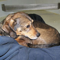 specific matings. At UNC, the hemophilia A dogs were originally Irish setters; the hemophilia B dogs were Cairn terrier/Beagle crosses; and the VWD dogs were all Scottish terriers. “Now all have been outbred for logistical reasons, to exclude unwanted traits that occur in different strains,” Nichols says.
“All of our breeding is for a purpose—to maintain the germ lines. Researchers design their experiments with planned breeding in mind, to have enough dogs for their studies,” say Nichols. Pregnant dogs with hemophilia receive prenatal care similar to pregnant women—factor levels are measured before labor and delivery, and product is given to prevent complications. “We transfuse the mother dogs weeks before delivery and continue until their postpartum bleeding stops,” Nichols says.
The blood for the hemophilic dogs’ transfusions comes from a separate colony of blood banking dogs. Most are large breeds with easily accessible veins, such as hound dogs or greyhounds. Like human blood, the donor dogs’ blood is typed. “We have about eight dogs that are donors,” says Lillicrap. “They have their blood taken every week or two.”
Canine Care
The dogs at both colonies are cared for by trained veterinary technicians, some of whom have worked with them for more than 25 years. “The people who work here love animals and are extremely knowledgeable about them,” says Nichols. At UNC, the dogs have indoor/outdoor runs and an acre of outdoor play area to romp in. “They’re not kept in cages, but in runs that are cleaner than our hospital,” says Roberts, partially joking. “They’re out in paradise next to University Lake.”
The dogs are socialized with each other and receive plenty of human contact. Just like family pets, they are taught commands and receive treats as rewards. “The dogs are in the lab all the time with people,” says Nichols. Since they stay there for years, they become more than just lab animals. “They become companion animals,” says Lillicrap. “They are very well looked after and nurtured.”
The dogs have names. At the Canadian facility, litter mates receive names starting with the same letter of the alphabet—Mindy and Matteo, for instance are siblings. At the UNC lab, the dogs are named for personality or looks. “One of our inhibitor dogs, Cliffy (named for Clifford the Big Red Dog), is the lab prince right now.” One of the vet techs is a dog groomer and gives Cliffy’s black-and-white coat special attention. “He is groomed to look like he has a great big mustache,” Nichols says with a laugh.
Most of the dogs live out their lives at the labs, being used for research in middle and older age, Lillicrap says. “We don’t breed for a specific experiment and then euthanize the dog. We now have dogs that are 13 years old.”
Other Animal Models
Researchers cannot limit their animal subjects to only one species. Protocol requires them to progress from rodents, for instance, to larger animals, if the experiment is successful at each stage. Larger animals help researchers obtain the proper scale in terms of physiology and anatomy. Eventually, experiments are approved for human studies, called clinical trials. (See “Clinical Trials 101.”) This progression from one animal model to another and eventually to humans can take years.
Michele Calos, PhD, professor in the genetics department at the Stanford University School of Medicine in California, is a bleeding disorders researcher who uses a variety of lab animals. “A huge amount of work has been done on mice throughout genetics because they are less expensive, have a shorter life span and you can do experiments with such larger numbers,” she says.
Using mice helps researchers build confidence about treatments and techniques, but they have limitations. Their size and blood volume are two factors; so is their genetics. “For most, if not all, gene therapies, the delivery is a barrier,” says Calos. Even with the sometimes-successful delivery in mice, however, the results do not translate well. “You cannot go from a mouse model to humans,” she says. “It’s too risky.”
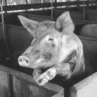
The intermediary can take the form of anything from pigs to primates. The UNC lab has housed pigs with severe type 3 VWD since 1942. Recent experiments on them with a type of interleukin show promise in elevating von Willebrand factor. The pigs helped researchers determine the proper dosing for humans. Calos and her colleagues tried pigs at first, because of their size. The clinical equipment for the procedure—the catheters and interventional radiology apparatus to inject the material into a liver vein—are the same for pigs and human patients. “The study in animals, piloting what we’re planning to do in humans later, was very useful to validate the approach down to the details.” But the hog model also had its hang-ups. The liver has a different structure and anatomy from humans. So the next step is to experiment on primates.
Primates are an ideal animal model primarily for one reason—their DNA. The genome of the rhesus monkey, a common lab animal, was sequenced in 2007; 93% of its DNA is identical to humans. “Some primate species have as much as 95% to 98% identical DNA as humans,” Calos says. “By going to baboons, we will be able to get something very similar to humans in the size, anatomy and genome,” she says. Since the baboon liver is similar to humans, Calos will be able to deliver the FIX gene therapy through a specific blood vessel to a particular lobe. “Most primates will tolerate human factor because it’s very similar to their own,” she says. “If the studies are successful, it’s a very strong prediction for success in humans.”
Assessing Animal Alternatives
With technological advancements occurring at a rapid pace, some might wonder why computer models or other techniques haven’t made animal experiments obsolete. “Animal models are still needed. I don’t believe a computer model can predict a dog’s response to a specific bleeding situation when given replacement therapy, for example,” says Roberts. Calos agrees. “There are so many factors involved, like the gene therapy delivery of DNA into the cells of functioning organs, which are only present in the animals.”
Cell culture studies, while useful, Calos says, have problems. The delivery actually worked better in the animals than the cell studies had predicted. “There is absolutely nothing in a computer model that would have given us this information,” Calos concludes.
Researchers try to use the minimum number of animals for an experiment, for ethical and practical purposes. Maintaining lab animals is expensive and requires expert care. “None of us would use animals if there were alternatives,” says Calos. “But there simply is nothing even close.”
National Resource
While none of the research animals has become a household name—no Lassies, Wilburs or Stuart Littles—together they have achieved their own notoriety. Researchers are indebted to all of them, but especially to the unique canine colonies at UNC-Chapel Hill and Queen’s University in Canada. “They have contributed enough knowledge about human hemophilia to make them an absolutely precious model,” says Roberts. “These dogs are a national resource.”
Animal Welfare
Lab animal facilities must meet stringent guidelines. In the US, they are overseen by several agencies, including the Association for Assessment and Accreditation of Laboratory Animal Care (AAALAC) International, a voluntary accreditation agency that provides peer review to hundreds of universities and other institutions; and the United States Department of Agriculture (USDA), which sets the standards by enforcing the Animal Welfare Act. The USDA can close a facility that is noncompliant. The UNC dog colony meets and exceeds USDA and AAALAC standards; the Canadian dog colony meets those set by the Canadian Council on Animal Care (CCAC), the national regulatory agency that makes onsite visits.
 In addition, universities must assemble an Institutional Animal Care and Use Committee (IACUC), comprising faculty, veterinarians and people from the community. The group reviews research studies and determines which ones receive approval.
Periodic inspections ensure that the facilities provide the animals with adequate food and water, housing, medical care and enrichment. The Animal Welfare Act standards stress that measures must be taken to avoid pain and suffering in the animals.
Social animals are required to have interaction with other members of their species, such as through communal housing. Enriching their environment is important to decrease distress and increase behaviors they display in the wild, such as foraging and social grooming. Examples include supplying rodents with nesting material and shelters in which they can hide or sleep. Running wheels provide exercise for them. Elevated resting areas are important for primates, which make their nests in tree tops. Hiding food in containers that must be manipulated to open keeps monkeys mentally engaged. Providing windows and mirrors allows primates to observe other animals and humans. Chew toys for dogs and hogs help prevent boredom.
The Animal Welfare Act further stipulates that laboratory dogs be given at least 30 minutes of exercise daily. It also recommends daily physical contact with people, including petting and playing.
To Learn More…
- Association for Assessment and Accreditation of Laboratory Animal Care (AAALAC) International
- Animal Welfare Institute
- Canadian Council on Animal Care
- Read the FDA regulations on genetically engineered animals
- Kulpa-Eddy JA, Taylor S, Adams KM. USDA perspective on environmental enrichment for animals. Institute for Laboratory Animal Research Journal 2005; 46:83–94.
- The Office of Laboratory Animal Welfare within the National Institutes of Health’s Office of Extramural Research establishes policies and publishes guidelines on the use of laboratory animals.
- The USDA’s Animal Welfare Information Center